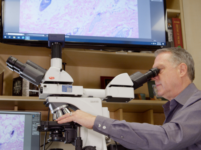
Dermatophyte Testing
Dermatophytosis (ringworm) is a fungal infection of the hair, superficial skin, and occasionally nails. It’s not caused by a worm and often lesions are not in rings. Dermatophytosis is instead caused by infection with one of three species of fungal organism: Microsporum canis, Microsporum gypseum, and Trichophyton mentagrophytes.
Common sources of infection include infected pets, rodents, contaminated environments, and occasionally the soil. While less common than many other skin diseases, dermatophytosis is a contagious and zoonotic disease that can in some cases be transmitted to humans. This makes it an important disease to assess and diagnose when the index of suspicion is high. Methods for diagnosing dermatophytosis include fungal culture, PCR testing and on occasion skin biopsy. In some cases, fungal spores may be identified on impression smear cytology raising the suspicion of dermatophytosis. Woods lamp examination may also increase suspicion of dermatophytosis.
If a veterinary dermatologist feels it is appropriate to test for dermatophytosis, most often he or she will collect hair and skin samples from the area(s) involved for one of these tests. Identification of the specific fungal organism responsible for skin lesions via fungal culture is the most reliable of these methods. Fungal cultures often take 14 to 21 days to complete to allow identifiable fungal organism growth.
When treating a pet for dermatophytosis it is imperative to treat the animal beyond clinical cure, when no further lesions can be seen. To confirm that the infection has been completely resolved a fungal culture is typically repeated to confirm successful therapy.

![Diagnostics - Biopsy 1 - Biopsy procedure Image 2 300dpi[1]-1](https://www.animaldermatology.com/hs-fs/hubfs/Diagnostics%20-%20Biopsy%201%20-%20Biopsy%20procedure%20Image%202%20300dpi%5B1%5D-1.jpg?width=800&height=600&name=Diagnostics%20-%20Biopsy%201%20-%20Biopsy%20procedure%20Image%202%20300dpi%5B1%5D-1.jpg)