Craig Griffin, DVM:
A very important part of understanding ear disease is realizing the difference between otitis externa and otitis media.
When you think about the ear, you have to realize that it's just skin. Skin everywhere on your body, you produce layers and layers of skin and debris, and it flakes off. In the ear, if things were to just come to the surface and sit there, the ear would fill up with debris. They push this against the eardrum, which can make it dilate or rupture. And once it ruptures, that can actually change that bone lining that middle ear. And at that point, it becomes a very difficult disease to treat. One of the key features of a normal ear is it cleans itself out, something called epithelial migration. And that self-cleaning, once it's damaged, means you're going to keep having recurrent ear infections. Even if you cleaned up the underlying disease in the original infection, that self-cleaning is what keeps the ear normal.
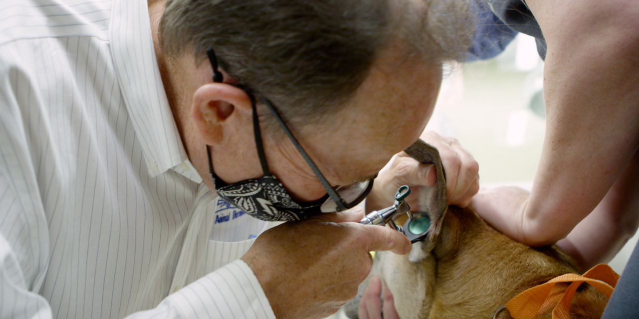
Video otoscopy is a tool that allows us to diagnose ear disease better. And basically, they have developed small cameras so I can now pass an otoscope down to the bottom of the ear canal here. There's a fiber optic light source going in, plus fibers that bring the image back from a lens that's literally at the bottom of the ear. So routine cleaning, you don't really know how well you've done it. You're cleaning, but you can't see what you're doing. You can't see the results. With video otoscopy, you can be sure you're cleaning the bad areas, you can be sure things as clean as you want. And then it's not just cleaning, I can see the pathology. I can treat certain areas of the pathology more specifically. It's just amazing to watch an animal that's been suffering for years, now all of a sudden no longer have chronic ear disease or recurrent ear infections, and it wants to play again.


-thumb-2.jpeg)